


Let’s be honest: external fixator complications happen. Pin site infections, loose frames, creeping malalignment — none of it is glamorous, but all of it is manageable with the right postoperative discipline. The good news? Most complications are largely preventable.
For manufacturers and distributors, understanding these risks isn’t academic — it directly shapes product design decisions. For clinical teams, systematic external fixation postoperative care is what separates smooth recoveries from drawn-out disasters.
This article is part of: External Fixators: Design, Types and Clinical Applications.
🗓️ Why Structured Postoperative Management Matters
Internal fixation is set-and-forget by comparison. External fixation demands active, ongoing involvement from patients, caregivers, and clinical teams, because the device sits half in and half out of the body. That reality creates four ongoing demands:
- Daily wound care at every pin site
- Constant vigilance for infection at the skin-pin interface
- Monitoring for frame loosening over time
- Managing patient compliance through weeks or months of wear
A structured postoperative protocol measurably reduces complication rates and improves outcomes. Most external fixator complications share one root cause: a break in the care routine. The American Academy of Orthopaedic Surgeons (AAOS) and international trauma societies publish the protocols that inform clinical practice — and they all agree on one thing: consistency beats intensity.
🧫 Pin Site Infection: The Leading External Fixator Complication
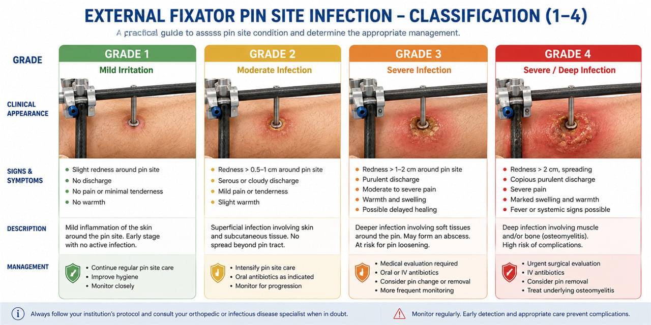
Pin site infection is the dominant external fixator complication, showing up in up to 20–30% of long-term fixation cases. The good news is that most infections are caught early, at a stage where hygiene and antibiotics resolve them.
Classifying External Fixator Complications: The Checketts-Otterburn Scale
The Checketts-Otterburn classification gives care teams a shared language for severity:
- Grade 1: Minor — redness and mild discharge; hygiene alone usually suffices
- Grade 2: Soft tissue infection — responds to oral antibiotics plus more frequent cleaning
- Grade 3: Deep soft tissue infection — IV antibiotics, possibly pin removal
- Grade 4: Bone infection (osteomyelitis) — rare but serious; demands aggressive management
Preventing External Fixator Complications: The Daily Pin Site Protocol
Effective external fixator pin site management starts at the OR table, not the bedside:
Surgical technique:
- No-burn drilling: low speed, sharp drill, pulsed advancement
- Minimize heat generation — bone necrosis is an infection portal
- Decompress skin around every pin (no skin tension)
- Never let trocar wires tent the skin
Daily pin site care:
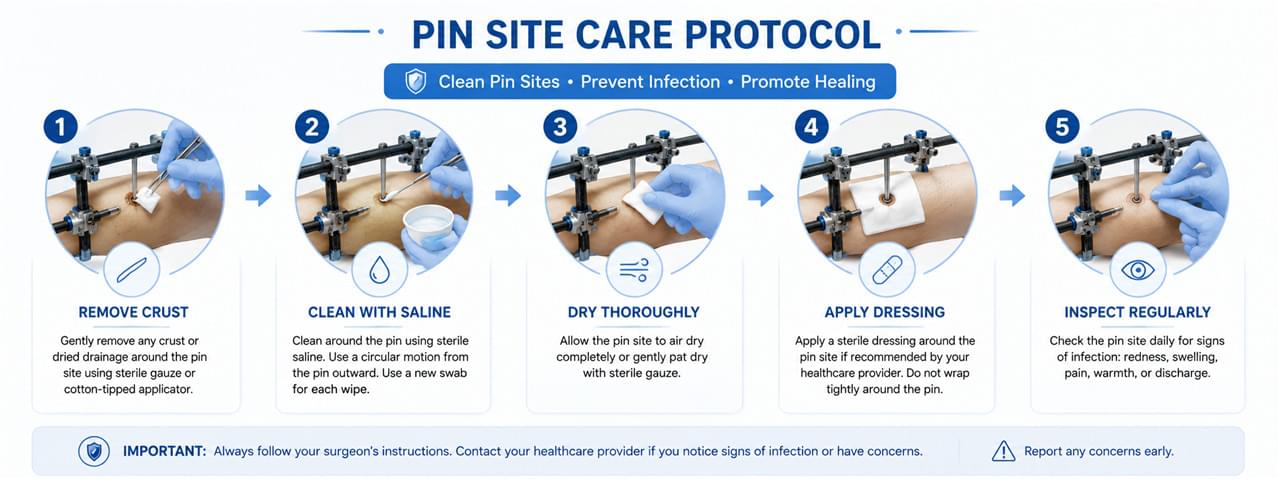
- Remove any crust formation (dry crust traps bacteria)
- Clean with standard saline or dilute chlorhexidine
- Pat dry
- Apply sterile dressing or leave open to air, per center preference
- Inspect for infection signs at every dressing change
Patient education:
- Demonstrate daily cleaning technique before discharge
- Teach the red flags of early infection
- Give clear contact instructions for concerns
One caveat worth knowing: pin site infection external fixator research hasn’t settled on a single best cleaning regimen. Even the Cochrane systematic review on pin site care — the highest evidence level in medicine — found no strong proof that one technique beats another. What the evidence does support: meticulous insertion technique and picking a protocol and sticking to it.
🔧 Frame Loosening and Management
Frame Loosening: A Silent External Fixator Complication
Loosening creeps up quietly — no alarm bells, just a frame that’s a little less solid than it was last week. Frame loosening is the second most common external fixator complication after pin site infection, and it compounds the first: a loose pin invites infection, and an infected pin loosens faster.
Causes:
- Micro-motion cycling causing bone resorption around pin threads
- Infection-driven bone resorption
- Inadequate bicortical purchase from the start
- Pin bending from excessive loading
Signs to check for:
- Pin site pain that worsens with activity
- Palpable pin movement on examination
- Radiolucency around pin threads on X-ray
Management:
- Replace loose pins (preferring adjacent sites)
- Re-tighten all frame connections
- Add extra pins if construct stability is compromised
📏 Malunion and Malalignment Risks
Malunion: The Creeping External Fixator Complication
External fixation permits gradual frame creep — a slow angular or rotational drift as the frame settles under load. It’s the complication nobody notices until the X-rays tell a different story.
Prevention:
- Weekly X-ray checks during the first 4 weeks for acute fractures
- Verify rotational alignment clinically at every visit
- Re-tighten all clamps at each follow-up
Managing progressive deformity:
- Early correction: frame adjustments under fluoroscopy
- Established malunion: may require re-operation
🦵 Knee and Ankle Joint Stiffness (Spanning Frames)
Frames that span the knee or ankle can quietly steal joint mobility. It’s an easy external fixator complication to overlook — it doesn’t threaten the limb, but it threatens function long after the frame comes off:
Prevention:
- Use spanning fixation only when clinically required
- Convert to non-spanning or internal fixation as soon as safe
- Start active range-of-motion exercises immediately after frame removal
Management:
- Supervised physiotherapy after removal
- Serial splinting for resistant contractures
- Rarely: manipulation under anesthesia
🦴 Refracture After Frame Removal
Remove the frame too early, and the bone may not yet have the callus density to handle normal loads:
Prevention:
- Confirm adequate union radiographically before removal
- Increase weight-bearing gradually
- Educate patients on refracture risk and precautions
Criteria for safe removal:
- Cortical bridging on 3 of 4 cortices on orthogonal X-rays
- No tenderness at the fracture site
- Patient can perform a one-leg stance on the affected limb
🏭 Manufacturer Implications: Design for Complication Reduction
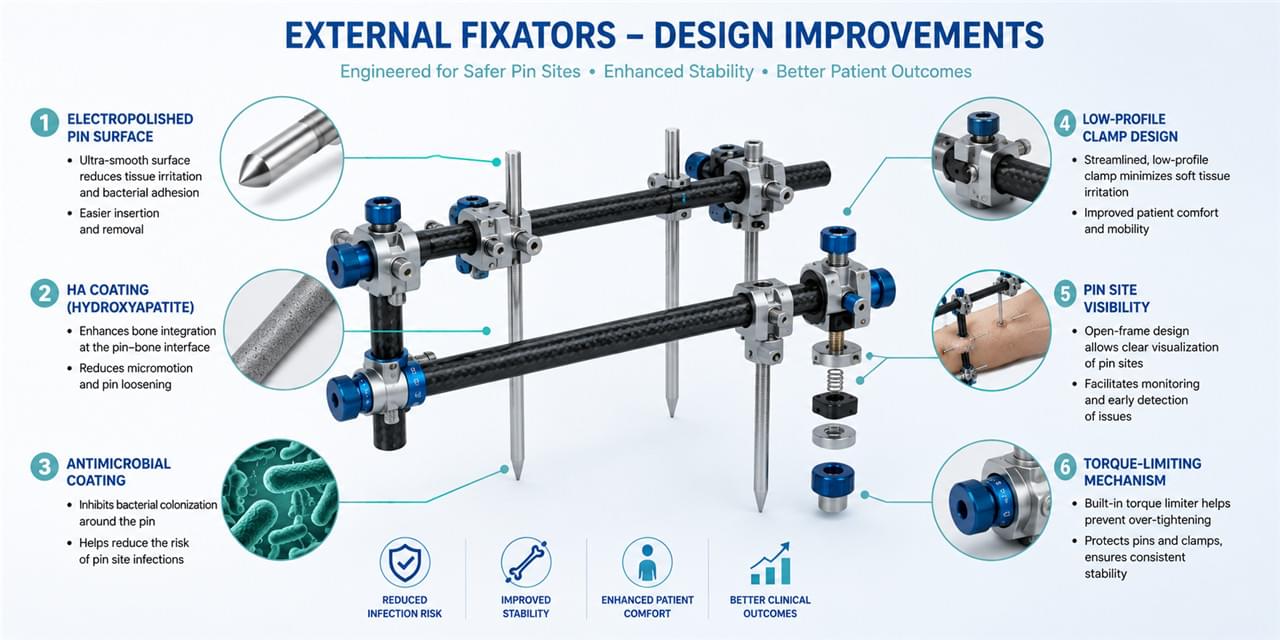
Understanding external fixator complications should sit at the center of device design priorities, not the periphery:
- Pin surface finish: electrolytically polished pins reduce bacterial adhesion at the skin interface
- HA-coated pins: emerging evidence for reduced loosening and infection
- Antimicrobial coatings: silver-doped or TiN options — an active product development area
- Low-profile clamp design: less skin pressure at clamp-skin interfaces
- Visual inspection access: frame geometry that allows pin site inspection without disassembly
- Torque-limiting assembly: color-coded or torque-limited clamps prevent over- and under-tightening
The evidence on coatings is still evolving — a recent systematic review and meta-analysis on pin coatings found no statistically significant infection-rate advantage for coated pins over stainless steel. That’s not a reason to ignore coatings, but it’s a reason to design for the whole system, not chase a single feature.
🛡️ Infection Prevention
The WHO guidelines on surgical site infection prevention provide the systemic context for infection prevention in orthopedic procedures, including external fixation.
Key systemic measures:
- Pre-operative antibiotic prophylaxis (standard for external fixation procedures)
- Sterile technique at pin insertion
- Perioperative glucose management in diabetic patients
- Pre-operative smoking cessation counseling
❓ FAQ: External Fixator Complications
Q1: How serious is pin site infection in external fixation?
Most cases are Grade 1–2 — minor, and resolved with aggressive local wound care plus oral antibiotics. Grades 3–4 are rare but serious, requiring IV antibiotics and possible pin removal. Deep bone infection (osteomyelitis) is the worst-case sequela.
Q2: Can infected pins be left in place?
Grade 1–2 infections are usually manageable without pin removal. Grades 3–4 generally require removal, with replacement at a different site if frame stability demands it.
Q3: How often should pin sites be cared for?
Daily care is the standard recommendation during active fixation. Some protocols allow twice-daily care in the early phase. The exact regimen varies by institution — what matters is consistency and compliance.
Q4: How is frame loosening detected early?
At each follow-up: palpate every pin for movement and tenderness, check X-rays for radiolucency around pins, and torque-test the clamps to catch loosening at connections.
Q5: What should patients do if they notice increased pin site redness?
Contact the treating team immediately. While waiting, increase cleaning to twice daily. Take documented oral antibiotics only on physician direction — no self-medicating with over-the-counter products.
🏁 Conclusion
External fixator complications — led by pin site infection and frame loosening — are eminently manageable with structured prevention and monitoring. For device manufacturers, complication reduction is a primary development objective: better pin surface finishing, thoughtful coatings, and ergonomic frame designs all translate into better clinical outcomes and sharper competitive positioning.
Return to: External Fixators: Design, Types and Clinical Applications.
Interested in pin surface treatment technologies or antimicrobial coating solutions for external fixation systems? Contact our team to discuss technical options.
⚠️ Medical Disclaimer
This article is for informational purposes only. All clinical management decisions must be made by qualified orthopedic professionals. Infection management protocols should follow institutional guidelines.
 English
 Arabic
 French
 German
 Italian
 Japanese
 Korean
 Portuguese
 Turkish
 Russian
 Spanish