


Complex fracture external fixation is one of the most technically demanding corners of orthopedic trauma surgery. High-energy mechanisms, mangled soft tissue, open wounds, and fracture patterns that look like a jigsaw puzzle — these are the cases where external fixation stops being an option and becomes the only sensible move.
This article walks through the clinical reasoning behind complex fracture external fixation using representative case scenarios. Each one illustrates a decision-making principle you can carry into the next trauma case — or the next procurement meeting. This article is part of: External Fixators: Design, Types and Clinical Applications.
🤔 What Makes a Fracture “Complex”?
Not every broken bone needs this level of firepower. Complex fracture external fixation becomes necessary when one or more of these factors show up:
- Open fracture (Gustilo Type II/III): significant soft tissue loss, contamination, or periosteal stripping — for the original grading criteria, see the landmark Gustilo and Anderson study that defined the classification every trauma surgeon still uses
- High-energy mechanism: motor vehicle accident, fall from height, gunshot wound — producing comminuted fracture patterns
- Periarticular location: fractures near joints with articular surface involvement
- Polytrauma context: a patient too physiologically unstable to tolerate prolonged anesthesia
- Vascular injury: associated arterial damage requiring vascular repair
- Infection or contamination: prior infection at the fracture site or heavily contaminated wounds
Spot any two of these in one patient, and you’re looking at a complex case by definition.
🦴 Scenario 1: High-Energy Open Tibial Fracture (Gustilo IIIB)
Clinical context: A 32-year-old motorcycle accident victim arrives with a comminuted mid-shaft tibial fracture, a 15cm laceration, significant muscle loss, and periosteal stripping. Classic Gustilo IIIB.
Why external fixation:
- The contaminated wound makes immediate IM nailing unsafe — implanting metal into a dirty field invites infection
- The wound needs serial debridements over 5–7 days before it’s clean
- Soft tissue reconstruction (flap coverage) is planned before any definitive internal fixation
External fixation strategy:
- Monolateral frame spanning the tibia
- Three half-pins above and three below the fracture (standard 4.0mm diameter)
- Aggressive wound debridement in the same surgical sitting
- Vacuum-assisted closure (VAC) dressing applied
The AO Surgery Reference’s principles of open fracture management lays out this exact sequence — classification, debridement timing, and why external fixation is preferred over internal hardware for severe wounds. Worth a read if you’re involved in system design or hospital protocols.
Timing to conversion:
- Once the wound is clean and covered (days 7–14): conversion to IM nailing if bone stock allows
- If delayed or impossible: definitive external fixation until consolidation
Frame design requirements:
- Radiolucent (carbon fiber) bar preferred for imaging follow-up
- 4.0mm stainless steel half-pins with electropolished surface to reduce infection risk
- Adequate bar-to-bone distance so dressings and wound care aren’t blocked
⚡ Scenario 2: Polytrauma — Damage Control Orthopedics
Clinical context: A 45-year-old in a high-speed collision. Closed bilateral femoral shaft fractures, closed head injury, hemodynamic instability. The OR clock isn’t ticking — it’s screaming.
The DCO principle: definitive fixation gets deferred. What the patient needs right now is rapid temporary stabilization to cut blood loss, reduce fat embolism risk, and dampen the inflammatory storm. The damage control orthopedics external fixator isn’t about elegance — it’s about speed and reversibility.
External fixation strategy:
- Bilateral monolateral femoral external fixators
- Applied in 15–20 minutes each under fluoroscopy
- Large-diameter (5.0–6.0mm) Schanz pins for adequate stability
- Spanning roughly 30–40cm proximal and distal to the fracture
Clinical outcome objective:
- Control hemorrhage and eliminate fracture motion
- Allow the ICU team to manage the patient without unstable limbs in the way
- Convert to IM nailing within 24–72 hours once physiology stabilizes
In external fixation of high-energy fractures with polytrauma, the frame is a bridge — not a destination.
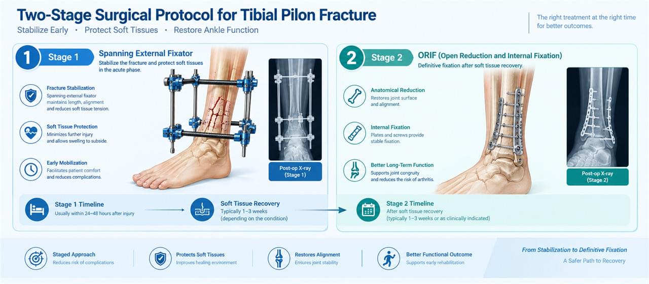
🦶 Scenario 3: Tibial Plafond (Pilon) Fracture
Clinical context: High-energy axial loading injury. Severely comminuted distal tibia articular fracture, significant swelling, fracture blisters developing. Operating on this today would be a disaster.
The “Fix and Flip” Protocol:
This well-established approach treats tibial plafond fractures in two deliberate stages, with external fixation as the bridge between them.
Stage 1 (immediate):
- Spanning external fixator across the ankle joint
- Fibula ORIF to restore length if the fibula is fractured
- The frame maintains length, alignment, and ankle position while soft tissues settle
Stage 2 (10–21 days later):
- Swelling resolved, skin condition improved, blisters gone
- Definitive ORIF of the tibial plafond
- External fixator removed
Frame requirements:
- Spanning configuration: pins in the tibia above the fracture plus two pins into the calcaneus or talus
- Maintains ankle neutral position and tibial length
- Radiolucent bar is essential for repeat CT planning
Periarticular fracture external fixation is usually a staged affair — patience with the soft tissues pays off in the final result.
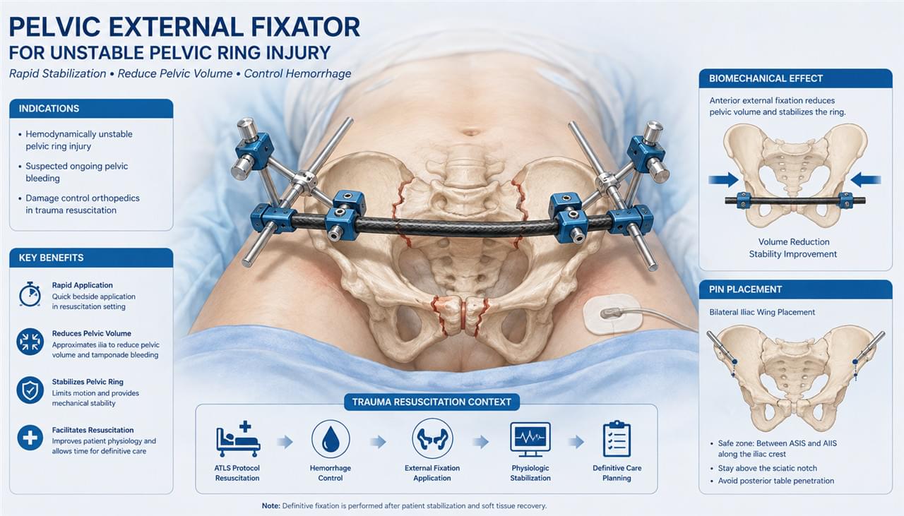
🛡️ Scenario 4: Complex Pelvic Ring Disruption
Clinical context: Unstable pelvic ring injury (Tile C, Young-Burgess APC III), hemodynamic instability, hemorrhagic shock. This patient is bleeding into the retroperitoneum and the clock is brutal.
External fixation as hemorrhage control:
- Anterior pelvic external fixator applied within 30 minutes in the trauma bay
- Pins into the anterior iliac crest bilaterally
- Reducing pelvic volume directly decreases retroperitoneal bleeding
Critical design requirements:
- Quick-connect system for rapid application in the trauma bay
- Standard 5.0mm or 6.0mm Schanz pins
- Simple two-pin bilateral configuration
- Pelvic fixator sets must be pre-positioned in trauma resuscitation areas — there’s no time to hunt for components
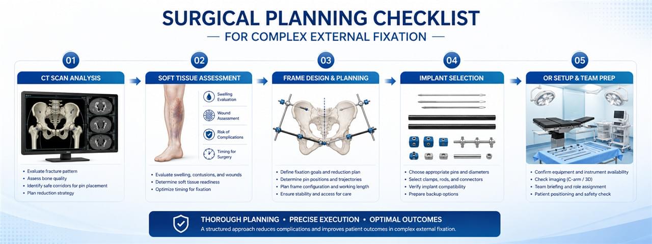
📐 Surgical Planning for Complex External Fixation
Successful complex fracture external fixation starts long before the first incision. Meticulous planning separates smooth cases from chaos:
The Planning Checklist for Complex Fracture External Fixation
- Fracture characterization: plain X-rays plus CT for fracture pattern and comminution assessment
- Soft tissue assessment: photo documentation and zone of injury mapping
- Frame configuration design: pin placement levels, bar configuration, spanning vs. non-spanning
- Implant selection: pin diameter, length, and material matched to patient size and bone quality
- Operating room setup: fluoroscopy positioning, traction table availability, backup internal fixation ready
The FDA’s guidance on orthopedic external fixation devices and AAOS clinical practice guidelines anchor current best practices — both worth bookmarking for procurement teams.
🏗️ Frame Configuration Strategies for Complex Cases
Bridging Frames in Complex Fracture External Fixation
The frame spans the fracture zone AND the adjacent joint:
- Maintains the joint in a functional position during healing
- Used for: wrist fractures, tibial plafond (ankle spanning), pilon fractures
- Risk: joint stiffness with prolonged application
Non-Bridging Frames in Complex Fracture External Fixation
The frame stabilizes the fracture WITHOUT crossing the adjacent joint:
- Allows early joint mobility during healing
- Preferred when fracture location permits non-spanning pin placement
- Better long-term functional outcomes when applicable
Delta Frame Configuration in Complex Fracture External Fixation
For added stability in tibial fractures:
- Standard monolateral pins plus an additional oblique pin from the distal fragment to the opposite cortex
- Creates triangular geometry that dramatically improves rotational stability
❓ FAQ: Complex Fracture External Fixation
Q1: How soon should external fixation be applied in an open fracture?
Definitive open fracture debridement — and external fixation if indicated — should happen within 6–8 hours of injury when possible. Provisional splinting and IV antibiotics start at the injury site. Earlier definitive treatment measurably reduces infection risk.
Q2: Can complex periarticular fractures be treated definitively with external fixation?
Yes, in selected cases — particularly elderly patients with poor bone quality, significant comorbidities, or when internal fixation can’t be performed safely. Functional outcomes are generally lower than internal fixation in young, active patients.
Q3: How long does swelling take to resolve before pilon fracture ORIF?
Typically 10–21 days. The clinical cues: return of skin wrinkles (the “wrinkle test”), resolution of fracture blisters, and normalization of compartment pressures. CT is performed at this stage for definitive preoperative planning.
Q4: What are the risks of prolonged complex fracture external fixation?
Pin site infection tops the list, followed by pin loosening, mal-union from gradual frame movement, joint stiffness with spanning frames, and a real psychological burden on the patient. Regular follow-up catches these early — that’s non-negotiable.
Q5: Is conversion from external fixation to IM nail safe?
Conversion within two weeks is generally safe with clean pin sites. Delayed conversion (beyond two weeks) carries significantly higher nail infection risk if pin sites are colonized. In delayed conversions, culture the sites and avoid nail contact with contaminated pin tracts.
🏁 Conclusion
Complex fracture external fixation demands surgical judgment, thoughtful frame design, and relentless postoperative management. For manufacturers and distributors, complex trauma is where demand for reliable, versatile systems really lives — radiolucent components, appropriately sized pins, and clamps that hold up under pressure.
Return to: External Fixators: Design, Types and Clinical Applications.
Looking for complete external fixation system solutions for complex trauma applications? Contact our team for product catalog and OEM capabilities.
⚠️ Medical Disclaimer
This article is for informational purposes only. Complex fracture management requires specialist orthopedic surgeon assessment and decision-making.
 English
 Arabic
 French
 German
 Italian
 Japanese
 Korean
 Portuguese
 Turkish
 Russian
 Spanish